Upper Thigh Anatomy - 3. Rectus femoris, vastus medialis, vastus lateralis and vastus intermedius. Small and deep muscles which mainly externally rotate the thigh at the hip joint and stabilize the pelvis. These muscles help us to allow the. There are five muscles in the anterior thigh compartment: It is the largest bone in the body and is the only bone in the upper leg.
Ebraheim's educational animated video describes muscle anatomy of the thigh. Small and deep muscles which mainly externally rotate the thigh at the hip joint and stabilize the pelvis. Like the forearm, the upper leg, or thigh, has a dense arrangement of many muscles. Rectus femoris muscle, one of the quadriceps muscles on the front of your thigh. There are five muscles in the anterior thigh compartment:
The Thigh Muscles Dummies from www.dummies.com Iliopsoas muscle, a hip flexor muscle that attaches to the upper thigh bone. Like the forearm, the upper leg, or thigh, has a dense arrangement of many muscles. The hamstrings are those three muscles that are located in the back of the thighs. The femur is known as a long bone. Pelvic & upper thigh anatomy. Sartorius, and the four quadriceps muscles; The hamstring portion of the adductor magnus has a similar action to these muscles, but is located in the medial thigh. Related links to external sites (from bing) these images are a random sampling from a bing search on the term vascular anatomy of the thigh.
Related posts of muscle anatomy of upper thigh anatomy muscle attachments.
• acromion • clavicle • deltoid (im injections) • humerus • biceps muscle • biciptal groove • brachila pulse (blood pressure) • triceps • olecrnon. Muscles play an important role in the. (there are four types of bone: Rectus femoris, vastus medialis, vastus lateralis and vastus intermedius. Along the upper portion of the thigh, just lateral to the gracilis, the adductor longus muscle is ranked as the most anterior of this group of thigh muscles upper thigh anatomy. In clinical anatomy the thigh muscles are divided into three groups: The upper legs consist of three main muscles: Abductors are located on the upper portion of the outside of your thighs and hips, anchoring above on the pelvis, and below at various points on your outside thigh. Upper leg anatomy and function the upper leg is often called the thigh. Each type of muscle tissue in the human body has a unique structure and a specific role. The hamstrings are those three muscles that are located in the back of the thighs. They have a lot to do with how your hips move. Anterior muscles extend your legs and flex your thighs.
Upper leg anatomy and function the upper leg is often called the thigh. Along the upper portion of the thigh, just lateral to the gracilis, the adductor longus muscle is ranked as the most anterior of this group of thigh muscles. Anterior muscles extend your legs and flex your thighs. Anatomy muscle attachments 12 photos of the anatomy muscle attachments anatomy muscle attachments, anatomy muscle attachments quiz, human anatomy muscle attachments, knee anatomy muscle attachments, shoulder anatomy muscle attachments, human muscles, anatomy muscle attachments, anatomy muscle attachments quiz, human. They have a lot to do with how your hips move.
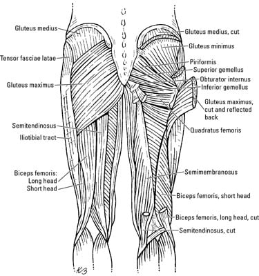
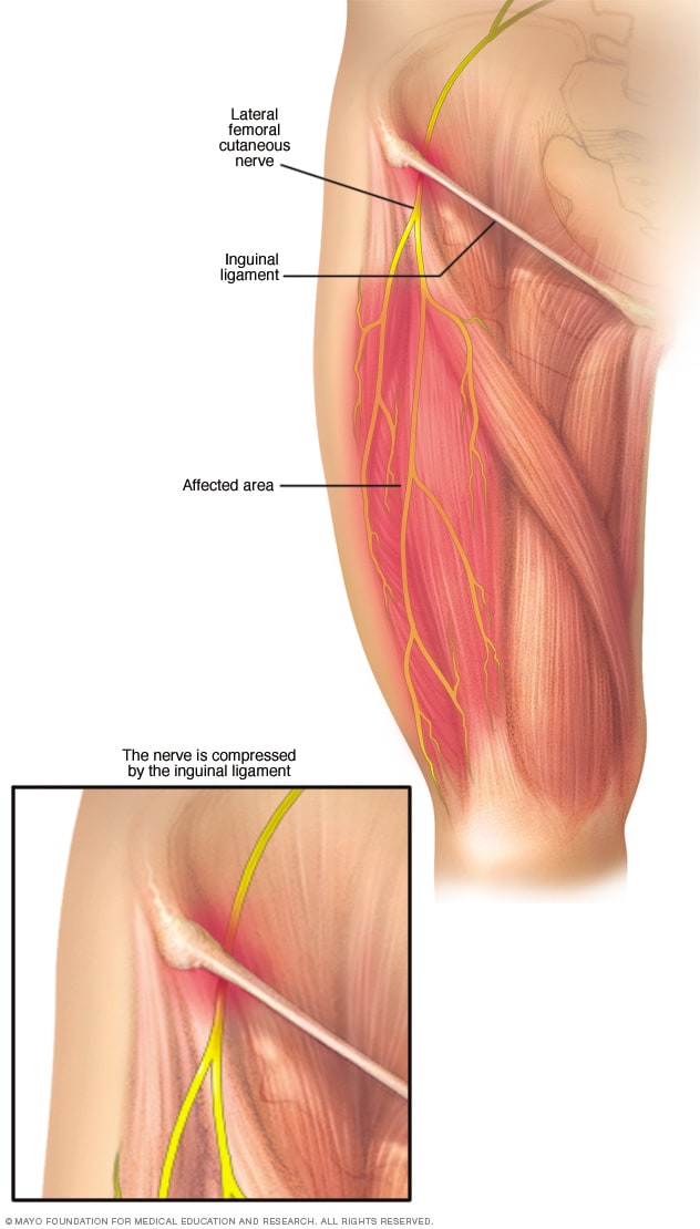
Meralgia Paresthetica Symptoms And Causes Mayo Clinic from www.mayoclinic.org Related posts of muscle anatomy of upper thigh anatomy muscle attachments. The anatomy of the legs can be divided into upper leg muscles and lower leg muscles. Upper leg anatomy and function the upper leg is often called the thigh. The femur is known as a long bone. Long bones, short bones, flat bones, and irregular bones.) • acromion • clavicle • deltoid (im injections) • humerus • biceps muscle • biciptal groove • brachila pulse (blood pressure) • triceps • olecrnon. Large and superficial muscles which mainly abduct and extend the thigh at the hip joint. The muscles located within the posterior compartment of the thigh are the biceps femoris, semitendinosus and semimembranosus.
The hamstring portion of the adductor magnus has a similar action to these muscles, but is located in the medial thigh.
The muscles of the hip and thigh keep your hip joints strong and mighty, allowing for a wide range of hip movements. Legs give us the freedom to run, walk, jump, climb, and negotiate the world around us. The thigh muscles don't just move your legs. Ebraheim's educational animated video describes muscle anatomy of the thigh. Like the adductors, the abductors are also responsible for stabilizing your knees during athletic and everyday movement. The muscles in the upper leg power many of our movements. 2, vastus medialis & intermedius muscles. In clinical anatomy the thigh muscles are divided into three groups: Rectus femoris, vastus medialis, vastus lateralis and vastus intermedius. These are the gluteus maximus, gluteus medius, gluteus minimus, and tensor fasciae latae. Abductors are located on the upper portion of the outside of your thighs and hips, anchoring above on the pelvis, and below at various points on your outside thigh. Lewis (1918) gray's anatomy 20th ed (in public domain at yahoo or bartleby) images: Upper leg anatomy and function the upper leg is often called the thigh.
The four muscles all extend the lower leg. It extends on to the base of the tail bone (sacrum), in front of the upper and lower anterior iliac spines, and on the front capsule (a bunch of three ligaments) of the hip joint. These are the gluteus maximus, gluteus medius, gluteus minimus, and tensor fasciae latae. Upper leg anatomy and function the upper leg is often called the thigh. Like the adductors, the abductors are also responsible for stabilizing your knees during athletic and everyday movement.
Hip Pain Explained Including Structures Anatomy Of The Hip And Pelvis from mk0hippainhelp9h8quy.kinstacdn.com Muscles play an important role in the. Like the adductors, the abductors are also responsible for stabilizing your knees during athletic and everyday movement. They have a lot to do with how your hips move. Lewis (1918) gray's anatomy 20th ed (in public domain at yahoo or bartleby) images: The four muscles all extend the lower leg. These muscles help us to allow the. One further muscle of the anterior knee is the small articularis genus muscle, it is occasionally is blended with vastus intermedius. Anterior muscles extend your legs and flex your thighs.
Each type of muscle tissue in the human body has a unique structure and a specific role.
Anatomy muscle attachments 12 photos of the anatomy muscle attachments anatomy muscle attachments, anatomy muscle attachments quiz, human anatomy muscle attachments, knee anatomy muscle attachments, shoulder anatomy muscle attachments, human muscles, anatomy muscle attachments, anatomy muscle attachments quiz, human. The hamstrings are those three muscles that are located in the back of the thighs. Related links to external sites (from bing) these images are a random sampling from a bing search on the term vascular anatomy of the thigh. The thigh is the area between the hip and the knee joint. Rectus femoris, vastus medialis, vastus lateralis and vastus intermedius. Medial muscles adduct and rotate your thigh, and posterior flex your leg and extend your thigh. Anatomically, it is part of the lower limb. The femur or thigh bone is one of the longest bones in the human body. Lewis (1918) gray's anatomy 20th ed (in public domain at yahoo or bartleby) images: Along the upper portion of the thigh, just lateral to the gracilis, the adductor longus muscle is ranked as the most anterior of this group of thigh muscles upper thigh anatomy. The muscles in the upper leg power many of our movements. Like the forearm, the upper leg, or thigh, has a dense arrangement of many muscles. Position the patient in supine position with feet pointing towards the magnet (feet first supine) position the patient over the spine coil and place the body coils over the thighs (anterior superior iliac spine down to knee joints) securely tighten the body coil using straps.

0 Response to "Upper Thigh Anatomy - 3"
Posting Komentar